
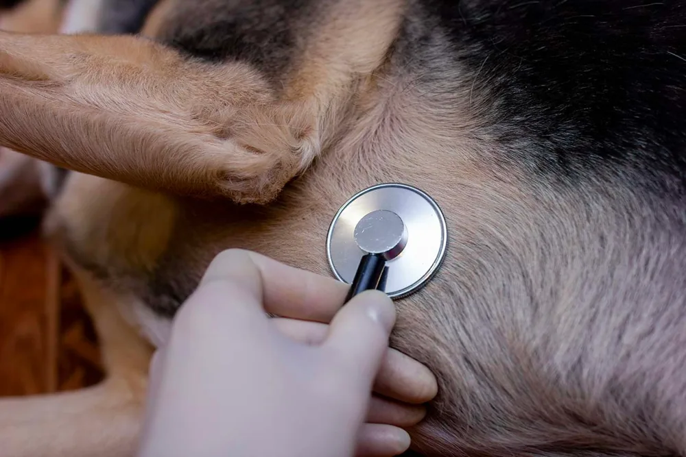
Subaortic Stenosis SAS is the acronym for Subaortic Stenosis which is tagged as a common genetic and congenital disease of the heart affecting specific dog breeds like German Shepherds along with Great Danes, Newfoundland, Boxers, Golden Retrievers, Bulldog,s and Rottweiler. Often times SAS is detected at an early age by hearing the heart murmur. The presence of Subaortic stenosis (SAS) in German shepherds or in any other canine breeds is usually established by noticing an abnormal heart sound. The sound is much like that of the rushing fluid (blood) through one of the heart valves. While the normal sound should be just lub-dub of the heartbeats, in case of the presence of Subaortic stenosis (SAS) a whistle of blood flow through a narrow blood vessel is heard.
Before delving deep into the disease there’s a need to understand the anatomy of the heart in simple words. The right chamber of the heart receives impure, deoxygenated blood from all organs and pumps it to the lungs for picking up fresh oxygen inhaled by the animal. The purified blood is received by the left chamber and it is responsible for channelizing it to all other body parts. Since the left chamber is the blood supplier its walls are thick and strong. The left ventricle or the pumping chamber pumps the blood to the aorta, which is the largest artery of the body. There’s a valve that separates these two units , it is named the aortic valve. The left ventricle narrows down to enter the aorta and this area is called the aortic outflow tract.
Mild, moderate or severe: the word that decides everything
SAS is not one condition with one outlook. Cornell divides it simply: “the narrowing can be mild, moderate, or severe” (retrieved 2026-08-12), and the difference between those words is the difference between a normal life and a serious risk.
Cardiologists grade it by how fast blood is moving through the narrowed area and the resulting pressure difference. The peer-reviewed review gives the cut-offs as equivocal at 2 to 2.5 m/s, mild at 2.5 to 3.5 m/s, moderate at 3.5 to 4.5 m/s and severe above 4.5 m/s, with corresponding pressure gradients (Subvalvular Aortic Stenosis review, retrieved 2026-08-12).
You do not need to memorise the numbers. You need to ask for them, because “he has a bit of a murmur” is not an answer you can plan around and “moderate, gradient 62” is.
What the severe end looks like. Cornell lists “weakness, breathing difficulty (dyspnea), fainting (syncope), and, in extreme cases, sudden death” (retrieved 2026-08-12).
Fainting deserves particular attention. A dog that collapses briefly during or just after exercise and then gets up as if nothing happened has done something that is easy to dismiss and should not be. Report it.
A heart problem your dog is born with
Subaortic stenosis, usually shortened to SAS, is not something a dog develops. It is a congenital defect, present from birth.
Cornell describes it as “a narrowing (stenosis) of the area underneath the aortic valve, that causes some degree of obstruction or blockage of the blood flow through the heart” (Cornell University College of Veterinary Medicine, Cardiology, retrieved 2026-08-12). The left side of the heart then has to push harder through a smaller opening, every beat, for life.
Cornell notes signs “may be present at birth (moderate or severe cases) or may appear in the first year of life”, and that SAS “most commonly occurs in large-breed dogs” (retrieved 2026-08-12).
A note on breed, since you searched for this one. Cornell does not single out the German Shepherd; it names large breeds as a group, and this is a large breed. A peer-reviewed review describes SAS as “the most common canine congenital heart disease” overall (Subvalvular Aortic Stenosis: Learning From Human and Canine Clinical Research, retrieved 2026-08-12). So this is worth knowing about as the owner of a large dog rather than as the owner of this specific breed.
How it is usually found: a murmur in a healthy puppy
Most owners meet SAS the same way. A vet listens to a bouncing, apparently healthy puppy at a vaccination appointment and hears a murmur.
Not every puppy murmur is SAS. Many are innocent and disappear as the puppy grows. But a murmur that persists past a few months, or one that is loud, is the reason your vet will suggest looking properly.
Looking properly means an echocardiogram. Cornell: “the degree of severity is assessed using several components of the ultrasound exam, especially Doppler ultrasound” (retrieved 2026-08-12). An X-ray shows the heart’s outline. Only ultrasound shows how fast blood is being forced through the narrowing, which is what severity actually means here.
Stenosis Means Narrowing
When the left ventricular tract lying just below the aortic valve has a scar-like narrowing (or stenosis) it leads to a condition that’s termed SAS. The area gets narrowed because of the presence of fibrous bands of tissues which are abnormal. Either these fibrous tissues are present from birth (congenital) or have an early development in the post-natal period.
The narrowing makes the blood-pumping process difficult; the left ventricle needs to exert greater pressure to pump blood through the narrowed passage. The blood forces its way out in the form of squirts and hence great turbulence is created within the heart chamber. This turbulence can be heard as a heart murmur on the left part of the chest.
Symptoms of SAS in German Shepherds?
When the vet hears the murmur in dogs, he or she becomes apprehensive of SAS in the animal. But it is not a definitive sign because there are many other clinical signs. In young puppies the stenosis is small (Grade I or II) but within the first 4-6 months of growth the stenosis enlarges and the heart murmur becomes more and more apparent. Puppies affected with SAS should be reevaluated when they are a year old in order to study and analyze the changes in cardiovascular status. Some other SAS symptoms are:
- Getting easily tired
- Coughing
- Spells of fainting (Syncope)
- Blue-tinged gums on body exertion
- SAS in a German Shepherd Dog Can be Severe
In SAS patients gradually there is a thickening of the walls of the left ventricle and there is also an enlargement of the ventricle because it needs to put in tremendous pressure to drive blood. This abnormal aortic valve hampers the heart’s electrical conduction and there is a disruption of its electric rhythm (or electrical arrhythmia). The pumping problem and the electrical disruption might even turn fatal. Hardly SAS patients outlive their third birthday. The Milder case of SAS in GSD or in other canine breeds is not considered a major medical issue, and the pet can enjoy a normal life span, with a slightly controlled lifestyle.
Radiograph Helps Diagnose SAS
The chance of heart failure can be identified with a chest radiograph. The clinical report shows dilation of the aortic valve (high-pressure squirting of blood is responsible for it). But the ideal test for SAS is ultrasound or echocardiography where the vet can measure the chamber size of the heart and the thickness of the wall. They study the correlation of the severity of stenosis with the pressure gradient.
The Ideal SAS Treatment
Beta-blockers (like atenolol) are the most effective drugs for treating SAS with the ultimate aim to normalize the exercising ability of the heart and help the animal enjoy a normal life span. The heart’s beta receptors that respond to epinephrine causes the heart to speed up its rate when exercising. The racing pulse leads to arrhythmia and frequent bouts of fainting. Beta-blockers are helpful in reducing the oxygen requirement of the heart. Surgery is a rare option; if it happens the surgeons excise the scar narrowing the passage. Balloon Valvuloplasty is another treatment for SAS that breaks down the scar and also dilates the stenosis.
Treatment: managing a plumbing problem with medication
Cornell: “the most common form of treatment is a medication given orally called beta blockers” (Cornell Cardiology, retrieved 2026-08-12). The peer-reviewed review notes that atenolol, being cardio-selective, “tends to be the beta-blocker of choice” (SAS review, retrieved 2026-08-12).
Beta blockers do not widen the narrowing. They slow the heart, which gives the left ventricle more time to fill and improves blood supply to the heart muscle itself. It is management, not repair.
What about surgery? Cornell is measured: “several surgical procedures have been performed with variable success” (retrieved 2026-08-12). Balloon dilation has been tried, and one difficulty documented in the review is that the gradient can climb straight back after an initially good result. This is a conversation for a veterinary cardiologist, not a decision to push for.
On outlook, the review records a median survival time of 56 months for dogs treated with atenolol, and lists the complications to watch for as left-sided congestive heart failure, endocarditis, arrhythmias, exercise intolerance, syncope and sudden death (retrieved 2026-08-12).
Mild SAS: fine for the dog, not fine for breeding
This is the single most important paragraph on the page for anyone who breeds, and it is the one most likely to be quietly ignored.
Cornell states that “mild subaortic stenosis is of no consequence“, and in the same breath that “it is usually recommended that the dog not be bred” (Cornell Cardiology, retrieved 2026-08-12).
Both halves are true at once, and that is exactly what makes it awkward. The dog itself is fine. It will run, work and live normally. But it carries a congenital heart defect, and breeding from it risks producing puppies at the moderate or severe end, where the risk is sudden death in a young dog.
So if you are buying a puppy from a large breed, it is entirely reasonable to ask whether the parents have been heart-screened by a cardiologist, and what the findings were. A breeder who has screened will tell you plainly.
What else causes these signs
| What you see | Consider |
|---|---|
| Murmur found in a puppy or young dog, otherwise well | Subaortic stenosis or another congenital defect |
| Middle-aged or older dog, tiring, breathing faster at rest, cough | Dilated cardiomyopathy |
| Starts a walk keenly then runs out of energy, young dog, wasted muscle | Muscle disease, rare |
| Weakness with pale gums and a swollen belly, sudden | Internal bleeding, see hemangiosarcoma. Emergency |
The age at which the problem appears is the most useful clue. Congenital defects show up in the first year. Acquired heart disease turns up in middle age and beyond. Our German Shepherd care guide covers routine health checks at both ends of life.
Living with a dog that has SAS
For a mild case, the honest answer is that very little changes. Cornell calls mild SAS “of no consequence” for the dog itself (retrieved 2026-08-12). Your dog exercises, trains and lives normally, and the diagnosis mostly matters on paper.
For moderate and severe cases, a few habits are worth building.
- Give the medication every day, on time. Beta blockers work by holding the heart rate down. Missed doses undo that.
- Never stop a beta blocker abruptly without talking to your vet. Stopping suddenly can cause a rebound.
- Know what to report. Any fainting or near-fainting, new coughing, breathing faster at rest, or a sudden drop in stamina.
- Ask what exercise is appropriate. This is a question for your cardiologist and the answer depends on the gradient. Do not guess in either direction, since needless confinement is its own harm.
- Mention SAS before any procedure. Anaesthesia is managed differently in a dog with a fixed obstruction in the heart.
- Keep the recheck echoes. Severity can change as a young dog grows, so a grade from twelve months ago is not necessarily today’s grade.
One more thing that is easy to miss. If your dog has SAS, mention it if a vet ever proposes a dental procedure, because endocarditis, infection settling on the abnormal area of the heart, is one of the recognised complications. Ask whether antibiotic cover is appropriate.
Questions worth asking
- Is this an innocent puppy murmur or a structural problem? The answer comes from an echocardiogram, not a stethoscope.
- What is the velocity and the pressure gradient? Get the numbers, not just the adjective.
- Mild, moderate or severe? This drives everything that follows.
- Does my dog need medication now, or monitoring?
- How often should we re-scan? Especially while the dog is still growing.
- What exercise is safe? Ask for something concrete.
- Should this dog be bred? Cornell’s guidance is that even mild cases usually should not be (Cornell Cardiology, retrieved 2026-08-12). Ask directly and take the answer seriously.

 Von Willebrands Disease in German Shepherds: Symptoms, Causes, and Treatments